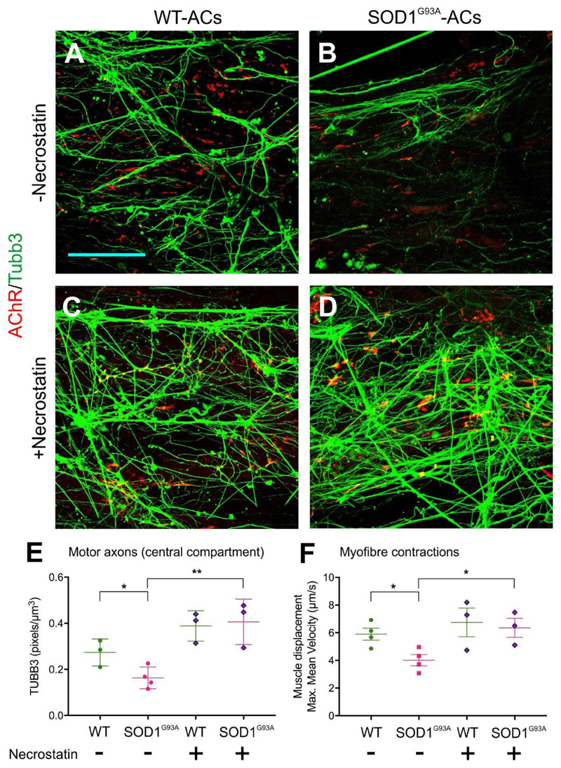
Figure 7.
Necrostatin rescues MN dysfunction caused by SOD1G93A-ACs. (A-D) Representative confocal images of the axons present in the myofiber compartment. ChR2-MNs were co-cultured with WT- or SOD1G93A-ACs in the absence or presence of Necrostatin. Scale bar 100μm (E) Axons are reduced in the myofibre compartment in MN co-cultures containing SOD1G93A-ACs compared to those containing WT-ACs. This decline was rescued by the addition of Necrostatin to the culture medium. (WT-AC: n=3; SOD1G93A-ACs: n=4; WT-AC +Necrostatin: n=3; SOD1G93A-ACs +Necrostatin: n=3 devices) Unpaired t-test, two-tailed p-value, *p<0.05; **p<0.01 (F) Myofibre contraction recorded in devices containing ChR2-MNs co-cultured with WT-ACs or SOD1G93A-ACs. Maximum mean displacement during light stimulation. We performed n=4 measurements (WT-ACs, SOD1G93A-ACs) or n=3 measurements (WT-ACs +Necrostatin, SOD1G93A-ACs +Necrostatin) with separate devices, each of them in triplicate. Unpaired t-test, two-tailed p-value, *p<0.05. Lines indicate mean and SD.

