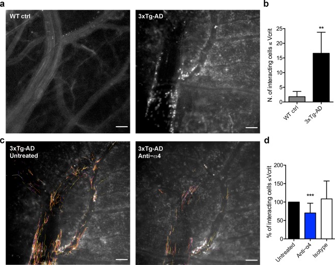
Figure 3.
Increased leukocyte–endothelial interactions in the brains of AD mice and the effect of α4 integrin blockade. (a) Representative images from EIVM experiments using a digital camera to record endogenous leukocytes labeled with Rhodamine 6 G, and automatic cell tracking in cerebral blood vessels of wild-type control (WT ctrl) and 3xTg-AD mice at 6 months of age. Interacting cells were automatically tracked in the field of acquisition based on their centroid fluorescence intensity. Scale bar = 100 µm. (b) Automatic Imaris analysis of endogenous leukocytes shows rare interacting cells in wild-type control (WT ctrl) mice compared to 3xTg-AD mice (**P < 0.01, Mann-Whitney U-test). (c) Representative images of leukocyte tracks in cerebral blood vessels of 3xTg-AD mice before (left panel, Untreated) and after (right panel, Anti-α4) i.v. injection into the lateral tail vein with a blocking antibody against α4 integrin. Scale bar = 100 µm. (d) Automatic Imaris analysis of leukocyte tracks before (Untreated) and after 30 min of treatment with the α4 integrin-specific blocking antibody shows a significant reduction in the percentage of interacting cells in the treated mice compared to the before-treatment control, set at 100% (***P < 0.001, Mann-Whitney U-test), whereas no effect was observed with an isotype control antibody. Bars show the percentage of interacting cells in the same venule over the course of 1 min. At least three vessels per mouse were analyzed and three animals were tested for each condition. Data are expressed as means ± SD (n = 3 mice (F) for all experimental conditions).

