Figure 1.
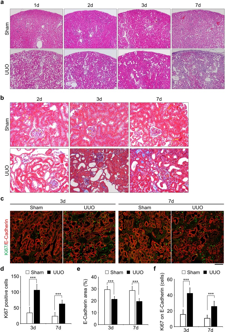
Epithelial tubular cells go into proliferative cycle upon obstruction of kidney. (a) Staining of Hematoxylin and Eosin (HE) of kidney slices obtained from various time points after obstruction. Sham-operated kidneys were used as a control. Images were taken at x100 magnification. (b) Masson’s trichrome staining. Fibrosis stained in blue. Images were taken at x400 magnification. (c) Co-immunostaining with Anti-Ki67 (green) and Anti-E-Cadherin (red). Ki67 was used for the identification of proliferative cells and E-Cadherin stained epithelial tubular cells. Bar, 100 µm. (d and e) Quantification of the number of Ki67 positive cells (d, n = 15) and E-Cadherin stained area (e, n = 15). (f) The number of Ki67 positive cells on E-Cadherin stained area (the number of Ki67 and E-Cadherin double stained area, n = 15). Data are as given averages ± SD (Standard Deviation); ***P < 0.001 (two-tailed unpaired Student’s t-test).

