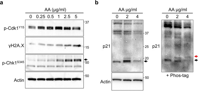
Figure 4.
p21 is involved in the response to the damage in vitro. (a) Immunoblotting with indicated antibodies of lysates from Aristolochic Acids (AA) treated HK-2 cells. AA was added into medium and cells were sampled after 24 h. Arrows indicate the height of intended bands. (b) Immunoblotting with anti-p21 of AA treated HK-2 cell lysates. Samples were separated in the presence (right) or absence (left) of 25 µM Phos-tag acrylamide containing SDS-PAGE gel. In the Phos-tag containing gel, the red and black arrows indicate the phosphorylated or non-phosphorylated p21, respectively.

