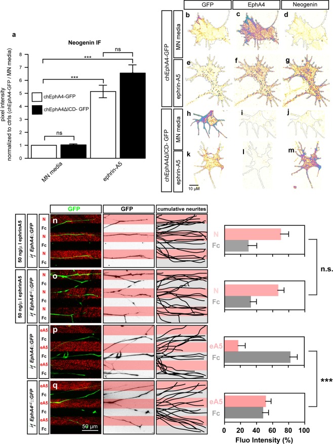
Figure 8.
The cytoplasmic tail of EphA4 is dispensable in potentiating the ephrin-A5 induced increase in Neogenin protein levels and sensitization of LMC axons to netrin-1. (a–m) LMC explants from chick spinal cords electroporated with either a chEphA4-GFP or chEphA4ΔICD-GFP expression plasmids were subject to a 20′ treatment of either MN media or ephrin-A5 at 100 ng/mL and immunostained for Neogenin (a) Quantification of Neogenin IF in growth cones shows that the ephrin-A5 induced increase in Neogenin signal occurs in growth cones expressing both plasmids (chEphA4-GFP: 5.1 ± 0.5-fold increase, p = 0.001, N = 3; chEphA4ΔICD-GFP: 6.6 ± 0.6-fold increase p < 0.001, N = 4). (b–m) Examples of GFP, EphA4 and Neogenin IF in growth cones quantified in (a). (n–q) Growth preference on protein stripes exhibited by LMC axons. Left panels: explanted LMC neurites expressing chEphA4-GFP and chEphA4ΔICD-GFP on netrin-1 (N)/Fc stripes bath treatment of ephrin-A5 (n,o) or ephrin-A5 (eA5)/Fc stripes (p,q). Middle panels: inverted images of GFP signals shown at left panels. Right panels: superimposed images of five explants from each experimental group representing the distribution of GFP+ LMC neurites. Quantification of lateral LMC neurites on first (pink) and second (pale) stripes expressed as a percentage of total GFP signals. Noted that both chEphA4-GFP and chEphA4ΔICD-GFP expressed LMC neurites show preferences over netrin-1 stripes. Minimal number of neurites: 85. Minimal number of explants: 13. N, netrin-1; eA5: ephrin-A5; error bars = SD; ***P < 0.001; statistical significance computed using Mann-Whitney U test.

