Figure 11.
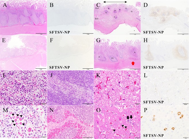
Hemophagocytosis and affected lymphatic nodules in the GI system of a fatal cat at 8 dpi with SFTSV and normal structures of surviving cat No. 1. (A,B) Normal aggregated lymphatic nodules in the lamina propria of the stomach of cat No. 1. SFTSV-NP-positive cells are not found. (C) Aggregated lymphatic nodules (*) in the lamina propria of the junction of pyloric gland region and duodenum. Decreased lymphocytes, necrotic germinal centers, congestion and missing nodule-associated epithelium (arrow) are seen in the lesions grossly identified as red spots in the stomach (in Fig. 8E). The villi are intact. St, stomach; Duo, duodenum; BrG, Brünner’s gland. (D) The lesional area was positive for SFTSV-NP. (E,F) Normal structure of the jejunum of cat No. 1. There are not found SFTSV-NP-positive cells. (G,H) Peyer’s patches in the jejunum. Necrotizing large sac-like lymphatic nodules (*) are positive for SFTSV-NP, while the internodular regions (IN) and villi are intact. Hemorrhaging occurred within the capsule of a nodule (red arrow). (I–L) Normal structures of surviving cat No. 1. I, sternal bone marrow; (J), mesenteric lymph node; (K,L), liver. (M) Hemophagocytosis in the sternal bone marrow from cat No. 2. Many hemophagocytic macrophages are present in the vascular space (arrowheads). (N) A mesenteric lymph node from cat No. 3. Numerous hemophagocytes are present in the medullary sinus. (O) Liver from cat No. 2. A large number of hemophagocytic macrophages (Kupffer cells, arrowheads) and round-shaped mononuclear cells (arrows) are seen in the sinusoids. (P) They are positive for SFTSV-NP (SFTSV-NP). The hepatocytes seem to be normal. (A,C,E,G,I–O); H&E staining, (C,D,F,H,P); SFTSV-NP, viral antigen by immunohistochemistry. Bars, (A–H), 1 mm, (I,K–M,O,P), 20 µm; (J,N), 100 µm.

