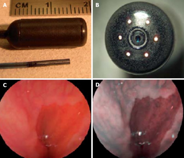
Figure 5.
Scanning single fiber endoscopy. A: Scanning fiber endoscopy (SFE) endoscope probes showing 9 mm rigid tip length of 1.2 mm diameter prototype and 18 mm capsule length of 6.4 mm diameter TCE. A front view of the distal end of the TCE is shown in (B) illustrating that the TCE is a standard SFE probe with collection fibers modified for capsule use. The gastroesophageal junction of a human subject is shown in single 500-line RGB image contrast (C) compared to postprocessed ESI contrast of the same SFE image frame (D). The lighter esophageal tissue is more clearly differentiated from the red-colored gastric mucosa in the ESI image. Reproduced with permission from Lee CM et al[46]. Copyright John Wiley and Sons, Inc.

