
FIGURE 2.
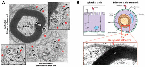
(A) Functional units of the adult human peripheral nerves, i.e., myelinated and nonmyelinated Schwann cell–axon units, depicting the distinct basal lamina that surrounds each Schwann cell–axon unit in situ. Red arrows indicate the basal lamina (BL) completely surrounding both myelinated (top inset) and nonmyelinated (bottom inset) Schwann cell–axon units. The Schwann cell membrane (M) is shown by black arrows. SC, Schwann cells; Ax, axons; MS, myelin sheath. (B) Sites of bacterial pathogens’ targets and entry into epithelia and peripheral nerves. Pathogenic bacteria enter epithelia at the apical side of the cells which anchor the basal lamina, whereas neurotrophic bacterial pathogens (e.g., M. leprae) must cross the basal lamina barrier, and thus attach to the basal lamina matrix proteins deposited around Schwann cell–axon units. The micrograph (adapted from reference 70) shows myelinated Schwann cell–axon units with the basal lamina (BL), Schwann cell membrane (SCM), and the axons ensheathed by the myelin sheath (MS).