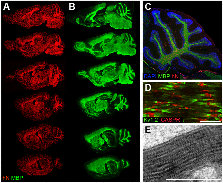
Figure 1. Engrafted hGPCs chimerize and myelinate the congenitally unmyelinated shiverer brain.
A-E, Myelination of congenitally hypomyelinated shiverer mice by human fetal tissue-derived glial progenitor cells. A-B. Representative sagittal images of an engrafted shi/shi × rag2−/− brain, sacrificed at 1 year of age. A, Human donor cells identified by an anti-human nuclear antibody (hN; red). B, Donor-derived myelin basic protein (MBP; green) in sections adjacent or nearly so to matched sections in A. All major white matter tracts heavily express MBP (which is all donor-derived in MBP-null shiverer mice). C, Sagittal view through cerebellum of a year-old engrafted shi/shi × rag2−/− brain. All cells were stained with DAPI (blue); donor cells were identified by human nuclear antigen (hN, red), and donor–derived myelin by MBP (green). D, Reconstituted nodes of Ranvier in the cervical spinal cord of a transplanted and rescued 1-year-old shi/shi × rag2−/− mouse, showing paranodal Caspr protein and juxtaparanodal potassium channel Kv1.2, symmetrically flanking each node. Untransplanted shiverer brains do not have organized nodes of Ranvier and, hence, cannot support saltatory conduction (Caspr, red; Kv1.2, green) E. Electron micrograph of a 16-week-old shiverer mouse implanted perinatally with hGPCs, shows a shiverer axon with a densely compacted myelin sheath.
Scale: D, 5 μm. E, 1 μm. Images from Windrem et al., 2008 (A-B); Goldman et al., 2012 (C-D); Windrem et al., 2004 (E); figure adapted from (Osorio and Goldman, 2016a).

