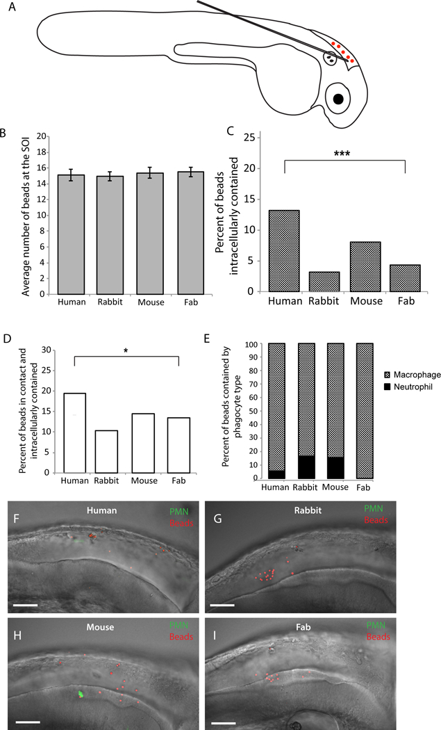
Fig. 1. Opsonizing polystyrene beads with human IgG increases bead-phagocyte interaction at SOI.

Prim25 stage mpx:EGFP transgenic zebrafish larvae (staged as described by Kimmel et al., 1995) with EGFP-expressing neutrophils and non-fluorescent macrophages (Renshaw et al., 2006) were injected into the hindbrain with polystyrene beads opsonized with human IgG, rabbit IgG, mouse IgG, and human IgG Fab fragment. (A) Microinjection of a 5 nL volume into the hindbrain ventricle through the otic vesicle was performed as described (Brothers and Wheeler, 2012), and inoculum was verified by screening larvae immediately post-injection. (B) Inoculum size within one hour post-injection is consistent among groups. (C-I) Images of the hindbrain ventricle were acquired via confocal microscopy at 4 hpi. Bead and phagocyte numbers were quantified based on 2 μm Z-stacks using a 40x/0.75 N.A. UPlanFL N objective. (C, D) A two-tailed Fisher’s exact test indicates a significant difference in the percentage of beads observed inside a phagocyte (C, p <.0001) and in contact with a phagocyte and intracellular (D, p < 0.05) between human IgG and human Fab opsonized groups. (E) Opsonized polystyrene beads are phagocytosed primarily by GFP-negative macrophages. Representative images reveal more bead-phagocyte interaction in fish injected with human IgG opsonized beads (F) as compared to beads opsonized with rabbit IgG (G), mouse IgG (H) and human Fab fragment (I). Images were acquired at 1024 × 600 pixels, and the number of slices for panels F, G, H, and I are 58, 53, 45, and 59, respectively. Movies including all of the slices are included as Supplemental Data Files M1E, M1F, M1G and M1H. Data are pooled from seven independent experiments. Although technical constraints limited cohort sizes in each experiment, reproducible differences were found from experiment to experiment. Number of fish scored; Human IgG n=18; Rabbit IgG n=17; Mouse IgG n=14; Human Fab fragment n=26. Scalebar = 50 μm.
