
Fig. 4.
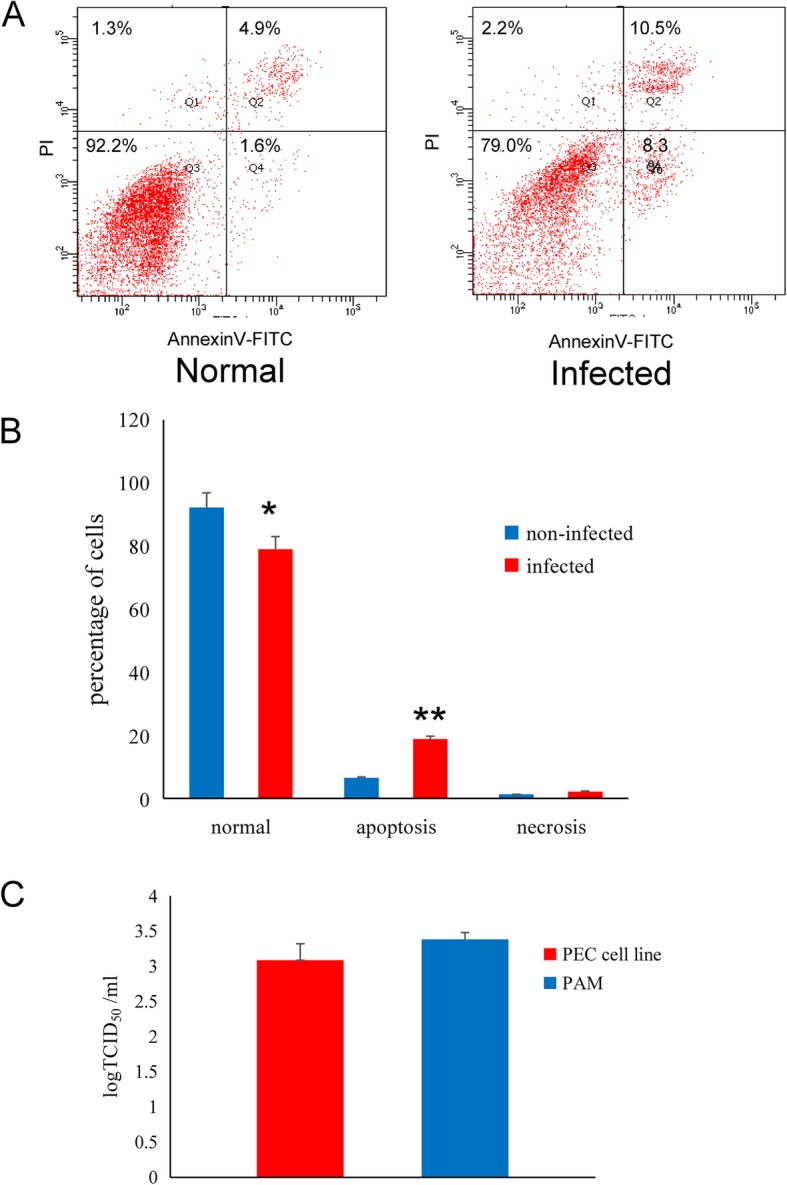
Susceptibility of PRRSV in the PEC cell line. a Comparison of apoptosis between normal and PRRSV-infected PEC cell lines. b A significant difference was observed between the normal and apoptotic cells after infected (* p < 0.05), the data were presented as Mean ± SEM and analysed by one-way analysis. c Comparison of PRRSV replication efficiency in the PEC cell line and PAMs (* p > 0.05), the data were presented as Mean ± SEM and analysed by one-way analyse