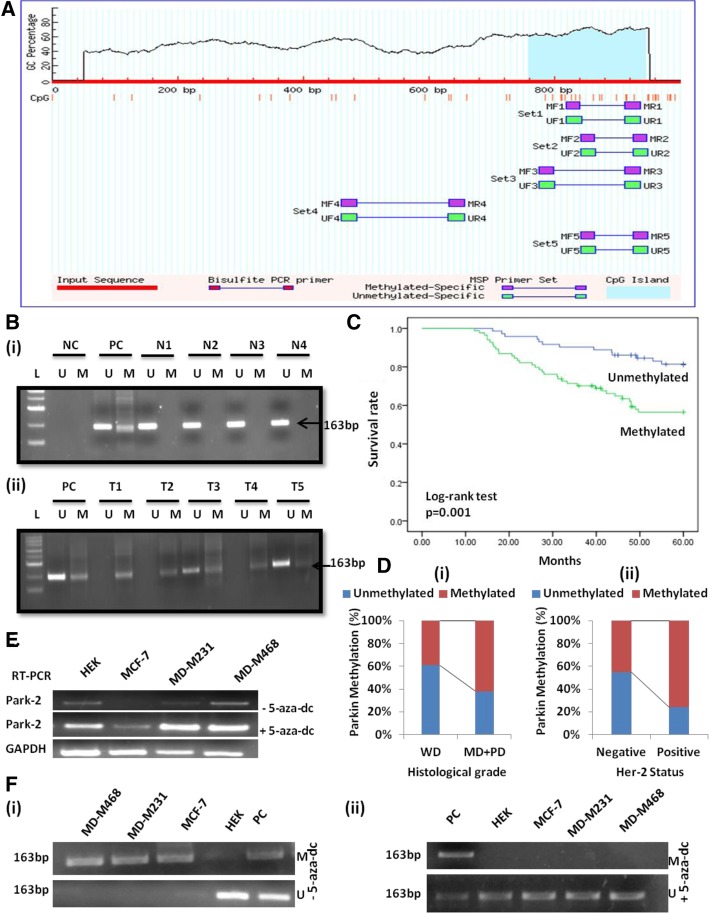
Fig. 1.
Parkin (PARK-2) methylation analysis. a Graphical representation of CpG islands (187) in the Parkin promoter region taken from MethPrimer (https://www.urogene.org/methprimer/); Criteria used: Island size > 100, GC Percent > 50.0, Obs/Exp > 0.60 b MS-PCR gel pictures, representing methylation of Parkin in (i) normal breast tissues & (ii) breast carcinoma tissues, PC-Positive control, NC-Negative control & L- Ladder (100 bp). c The survival curve was analyzed according to promoter methylation of Parkin protein (SPSS version 17.0). d Frequency distribution of Parkin methylation in (i) Histological grade (ii) Her-2 status (p < 0.05). Demethylating treatment with 5-aza-dC restored Parkin expression and unmethylated status in breast cancer cell lines. e Parkin mRNA expression through RT-PCR, showing that demethylating treatment with 5-aza-dC restored Parkin expression in MCF-7, MDA-MB-231, and MDA-MB-468 cell lines. HEK a non-tumor derived cell line was used as a positive control. GAPDH was amplified as an internal control. f Methylation-specific PCR of Parkin promoter in breast cancer cell lines MCF-7, MD-M231 & MD-M468 before (i) and after (ii) 5-aza-dC treatment. HEK was used as a positive control. PC- Positive control totally methylated and unmethylated bisulfite converted human genomic DNA

