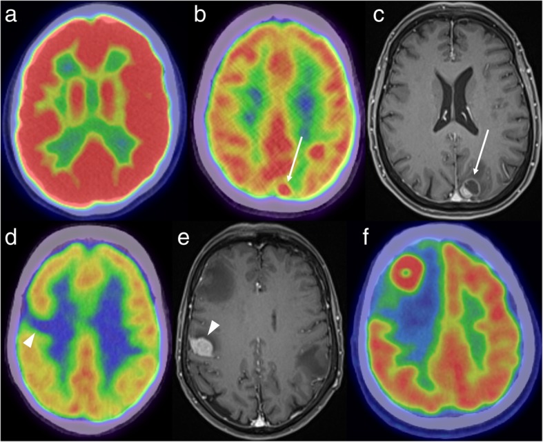
Fig. 1.

FDG-PET demonstrating normal high background uptake (a) - uptake is higher in the grey matter than in the white matter. A focus of high FDG uptake in the left parietal lobe (b, white arrow) corresponds to a mixed solid/cystic metastasis on the post-contrast MRI (c). An area of low uptake (d, white arrowhead) can also be due to a metastasis, as demonstrated on the corresponding MRI (e). FDG-PET in another patient (f) shows an FDG-avid mass in the right frontal lobe with surrounding photopaenia, consistent with oedema. Histology confirmed a solitary metastasis from a lung primary
