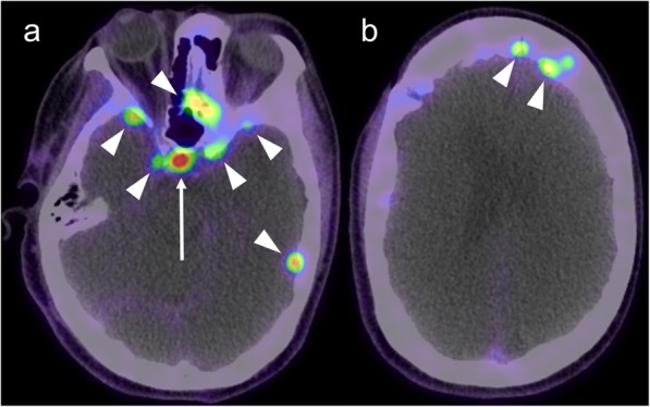
Fig. 13.

Post-contrast MRI (a) and GaTate-PET (b) in a patient with previous surgery for meningioma. A small enhancing nodule related to the falx cerebri (arrows) demonstrates GaTate-avidity, consistent with meningioma. In contrast, the more diffuse dural thickening (arrowheads) does not demonstrate GaTate uptake, and is thus consistent with post-operative change rather than en plaque meningioma
