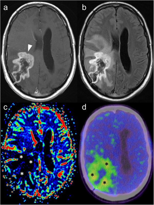
Fig. 8.

Post-contrast T1-weighted (a) and FLAIR (b) MRI images demonstrate an irregular peripherally-enhancing lesion in a patient with a known right temporo-parietal glioblastoma treated with temozolamide and radiotherapy. Given an absence of elevated cerebral blood volume on dynamic susceptibility contrast MRI perfusion (c), the possibility of pseudoprogression was raised. FET-PET (d) showed prominent tracer uptake, however, consistent with true tumour progression, which was confirmed histologically
