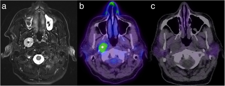
Fig. 9.

Axial T2 with fat saturation MRI (a) shows a mass in the right carotid space (asterisk), slowly enlarging on serial imaging (thus going against a metastasis). There is high uptake on FDG-PET (b), but no uptake on GaTate-PET (c), most consistent with a nerve sheath tumour (confirmed histologically)
