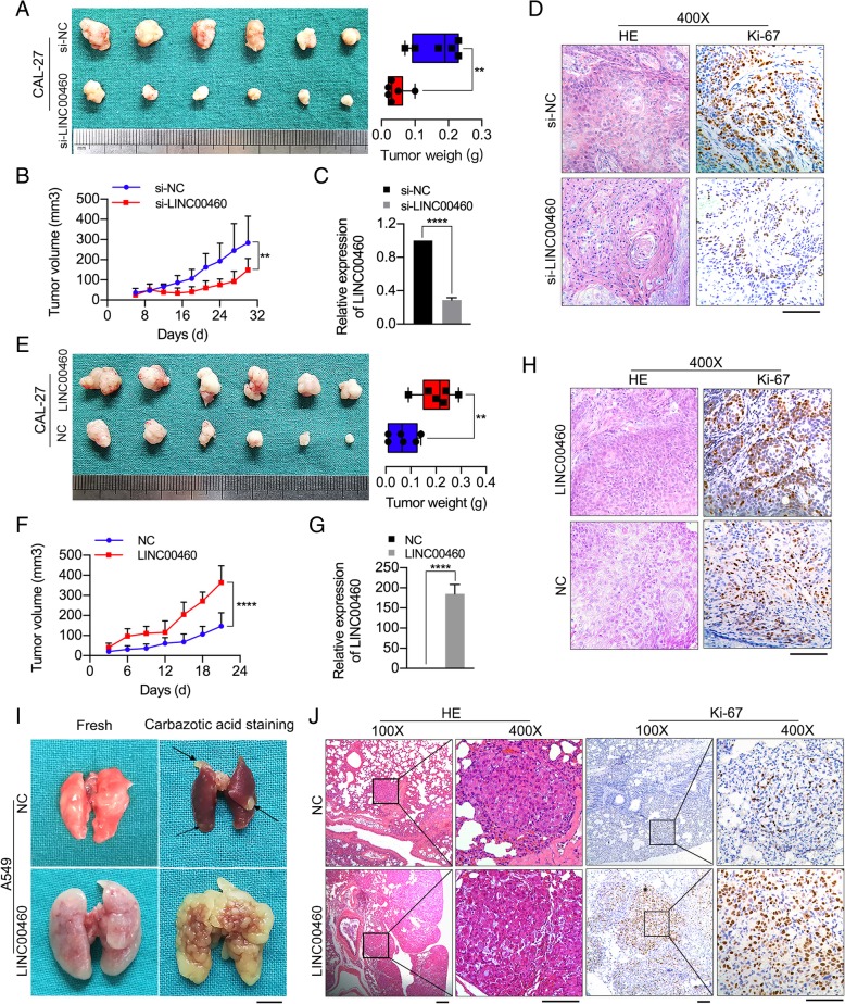
Fig. 3.
LINC00460 promoted HNSCC cell growth and metastasis in vivo. a The volumes and weights of tumors from CAL-27 tumor-bearing nude mice treated with cholesterol-conjugated si-LINC00460 or si-NC are shown n = 6/group, **p < 0.01. b The tumor growth curves of tumors from CAL-27 tumor-bearing nude mice treated with cholesterol-conjugated si-LINC00460 or si-NC are shown. **p < 0.01. c The levels of LINC00460 expression in tumor tissues formed from CAL-27 cells treated with si-LINC00460 and si-NC as determined by qRT-PCR. ****p < 0.0001. d H&E staining and IHC of Ki-67 in tumor tissues. Scale bar: 100 μm. e The volumes and weights of tumors in nude mice subcutaneously inoculated with CAL-27 cells stably transduced with LINC00460 at the end of the experiment are shown. n = 6/group, **p < 0.01. f The tumor volumes were calculated every 3 days for 3 weeks, and the tumor growth curves are shown. ****p < 0.0001. g The expression of LINC00460 in tumor tissues formed from CAL-27 cells stably transduced with LINC00460 was detected by qRT-PCR. ****p < 0.0001. h H&E staining and IHC of Ki-67 expression in tumor tissue. Scale bar: 100 μm. i Representative images of the lungs of mice inoculated with A549 cells stably transduced with LINC00460 or NC by tail vein injection for 8 weeks. The arrows show yellow nodules on the lung surfaces. Scale bar: 5 mm. j H&E staining and IHC of Ki-67 expression in lung tissues with tumor colonization. Scale bar: 50 μm

