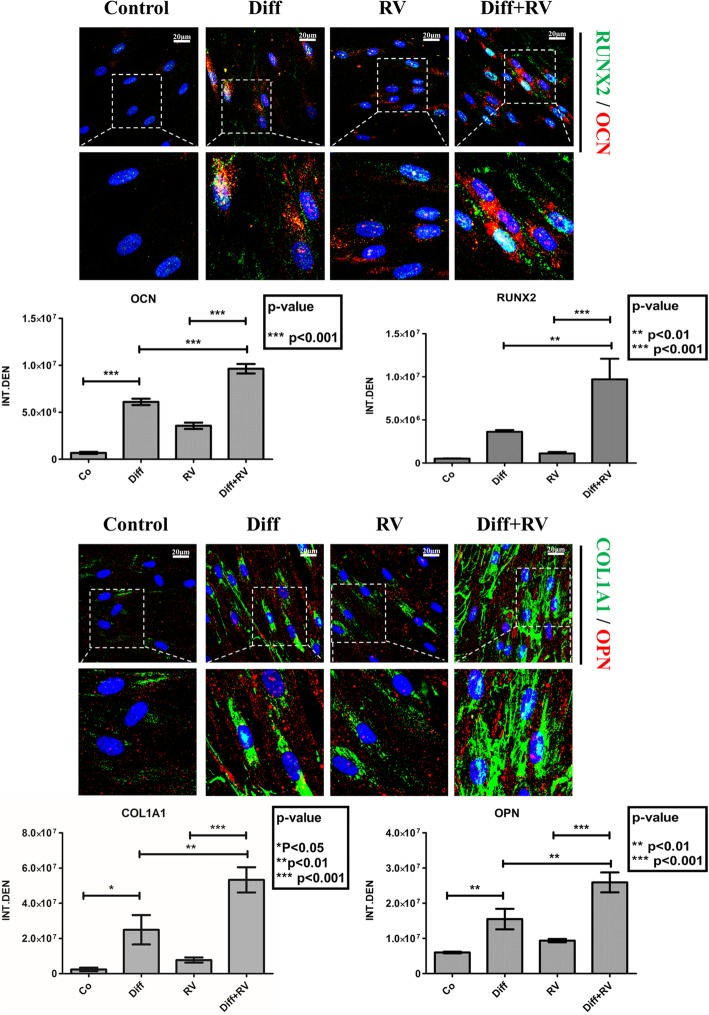
Fig. 5.
Immunofluorescence staining of osteoblastic differentiation markers in Human Gingival Mesenchymal Stem Cells. HGMSCs were plated on sterile coverslips, let adhere and cultured for 21 days in control medium or in differentiation medium (Diff) supplemented or not with 1 μM resveratrol (RV). At the end, the coverslips were fixed and processed for immunofluorescence staining of the osteogenic differentiation markers. Fluorescence staining was quantified with the ImageJ software

