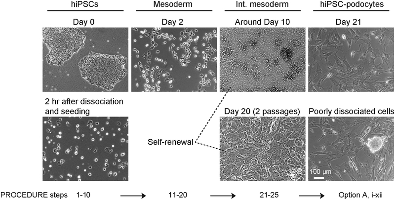
Figure 2|.
Morphological changes of human iPS cells at each stage of differentiation. Representative bright field images show human iPS cells before and after dissociation on day 0 when differentiation is initiated, mesoderm cells after 2 days of differentiation, intermediate mesoderm cells at around 10 days and after20 days of culture with two passages, as well as the terminally differentiated podocytes. An example of an image of podocytes derived from a population of cells containing a poorly dissociated colony (dotted red circle) is also shown. Int. mesoderm, intermediate mesoderm; hiPS-podocyte, human iPS cell-derived podocytes. Scale bar, 100 μm.

