Figure 5|.
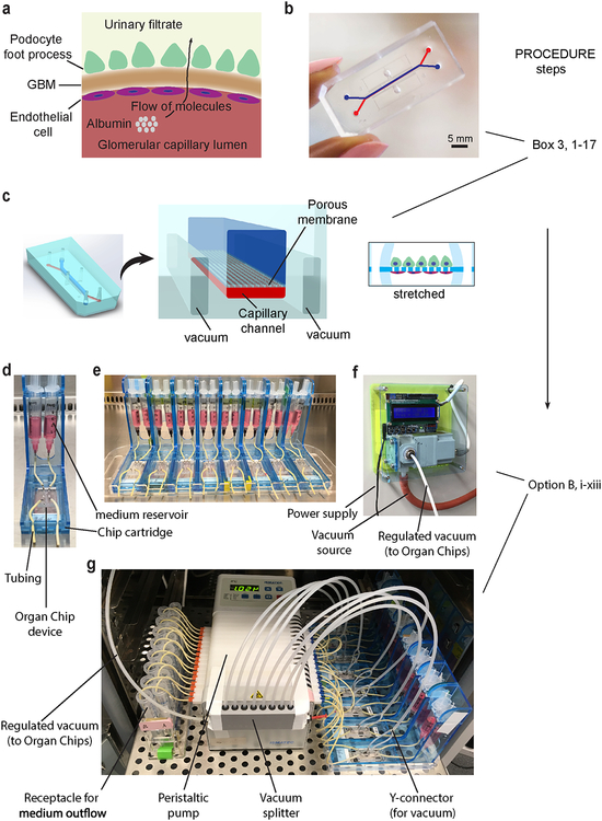
Design of microfluidic Organ Chip device to recapitulate the structure and function of the kidney glomerular capillary wall. (a) Schematic representation of the glomerular capillary wall showing podocyte and endothelial cell layers separated by the GBM to form capillary and urinary compartments. (b) Photograph of the microfluidic device engineered from PDMS. Scale bar, 5 mm. (c) Schematic representation of the microfluidic kidney Glomerulus Chip with microchannels replicating the urinary and capillary compartments of the glomerulus. The GBM is mimicked by using a porous and flexible PDMS membrane functionalized with the protein laminin. Cyclic mechanical strain was applied to cell layers by stretching the flexible PDMS membrane using vacuum. Example photographs of the experimental setup for Glomerulus Chip cultures show (d) a single Organ Chip microfluidic device connected to two reservoirs containing cell culture media for the urinary (right) and capillary (left) channels, (e) multiple Organ Chips placed on a cartridge built-in-house for handling of microfluidic devices, (f) photograph of the programmable vacuum regulator system built-in-house, (g) complete setup of the microfluidic cell culture system with the Glomerulus Chips connected to media reservoirs, inlet and outlet tubing, vacuum lines for stretching, and the peristaltic pump, as well as receptacles for media outflow. Figure modified with permission from Reference 5.

