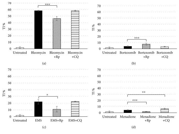
Figure 5.
DNA damage evaluated through the comet assay in the U937 cell line cotreated for 24 h with 0.1 μM rapamycin or 3 μM chloroquine and 67 μM bleomycin (a), 0.5 μM bortezomib (b), 1.9 mM ethyl methanesulphonate (c), and 1.3 μM menadione (d). Data are given in terms of percentage of DNA in the comet tail (tail intensity percentage: TI%). The error bars represent the standard deviation of two independent experiments. ∗ p < 0.05, ∗∗ p < 0.01, and ∗∗∗ p < 0.001.

