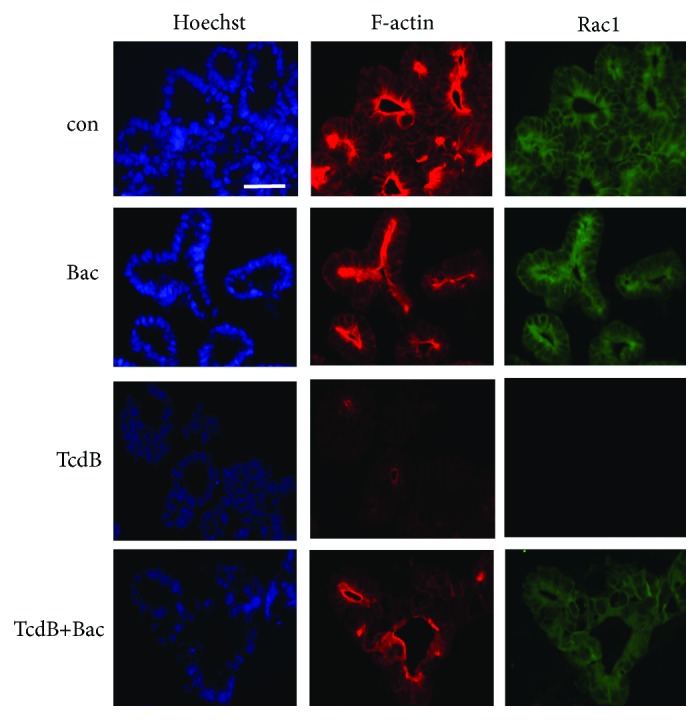
Figure 4.

Bac reduces the TcdB-induced glucosylation of Rac1 and destruction of F-actin in human intestinal organoids (“miniguts”). “Miniguts” were preincubated for 30 min at 37°C with or without 1 mM Bac. Then, TcdB (60 ng/ml) was added and the organoids were incubated for further 3 h. For control (con), cells were left untreated. After washing and extraction from Matrigel, “miniguts” were fixed and frozen sections were prepared. Nuclei, F-actin, and nonglucosylated Rac1 were specifically stained as described earlier [2, 3] and visualized by confocal fluorescence microscopy. Bar = 50 μm.
