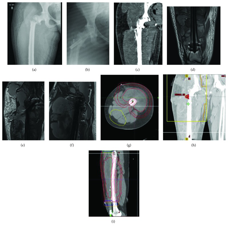
Figure 3.
(a) Preoperative AP pelvic X-ray showing a pathological fracture through the trochanteric region of the right proximal femur. (b) Postoperative AP pelvic X-ray showing stabilisation of the fracture using a CFR-PEEK cephalomedullary nail. (c) Postoperative coronal CT image with minimal scattering showing massive progression of the right thigh sarcoma. (d–f) Postoperative MR images showing progression of tumour mass with an increase in cystic components but no signs of postoperative infection. (g–i) CT planning images for radiotherapy with the absence of metal artefacts.

