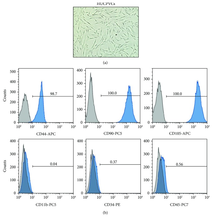
Figure 1.
Characterization of human umbilical cord perivascular cells (HUCPVCs). (a) Representative phase micrograph image of HUCPVCs in culture. HUCPVCs display a fibroblastic morphology (×100 original magnification). (b) Surface marker expression levels in HUCPVCs analyzed by flow cytometry. HUCPVCs highly express the stromal determinants CD44, CD90, and CD105 and are negative for CD11b, CD34, and CD45 markers. Solid blue histograms represent cells stained with fluorescent antibodies, and isotype-matched controls are overlaid in gray.

