Fig. 7.
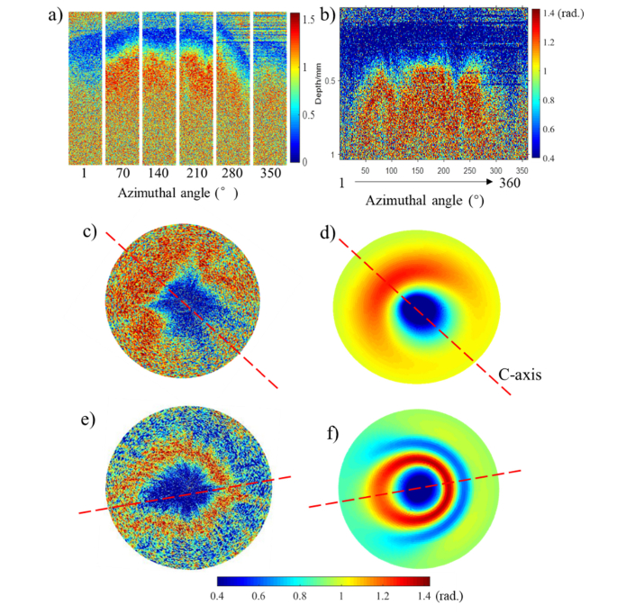
Real phase retardance images of human cervical tissue obtained by conical beam scan method and simulated retardance images using EJMC model. a): a series of B-scan retardance images in cervical center area acquired with a 45° incidence angle and a various of azimuthal angles. 360 A-scans of phase retardance extracted from the same data set are represented as a function of azimuthal angles in entire range of 1-360° with interval of 1° in conventional OCT image display format b) and polar format c). The corresponding simulated result is shown as d). e): 360 A-scans of phase retardance obtained in middle area represented as polar format. f): corresponding simulated result of e). Polar radius is 0.8 mm.

