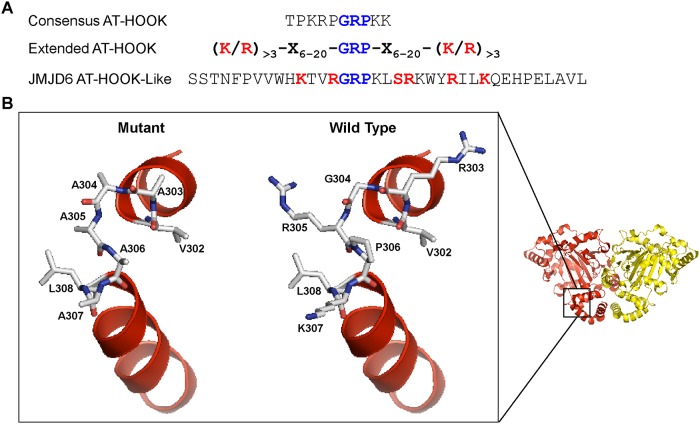
Fig 7. Predicted structure of the JMJD6 AT hook-like domain.
(A) Comparison of the amino acid sequences of a canonical AT hook, and extended AT hook, and the AT hook-like sequence of JMJD6. (B) PyMOL was used to visualize the JMJD6 dimer structure reported by Mantri et al (PDB ID 3K2O; [46]). The magnified area is a comparison between the predicted hinge area of the wildtype motif (right) and the motif with alanine substitutions of the GPRK amino acids at positions 303–307 (left).

