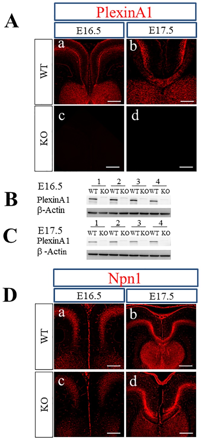
Fig 1. Localization of PlexinA1 and Npn1 in the coronal sections of WT and PlexinA1 KO brains at E16.5 and E17.5.

(A) Immunohistochemistry with anti-PlexinA1 antibody revealed the localization of PlexinA1 only in WT brains at E16.5 and E17.5 (a, b) as well as the lack of PlexinA1 in PlexinA1 KO brains (c, d). Scale bars: 200 μm. (B, C) Western blot analysis using anti-PlexinA1 antibodies was performed with tissue lysates from the medial regions covering the cingulate cortex, axons from the cingulate and neocortex, and the CC of E16.5 and E17.5 brains. PlexinA1 protein was detected in WT brains (B; E16.5, C; E17.5) but not in PlexinA1 KO brains (B; E16.5, C; E17.5). (D) Immunohistochemistry with anti-Npn1 antibodies revealed localization of Npn1 in both WT (a; E16.5, b; E17.5) and PlexinA1 KO (c; E16.5, d; E17.5) brains at E16.5 and E17.5. Scale bars: 200 μm.
