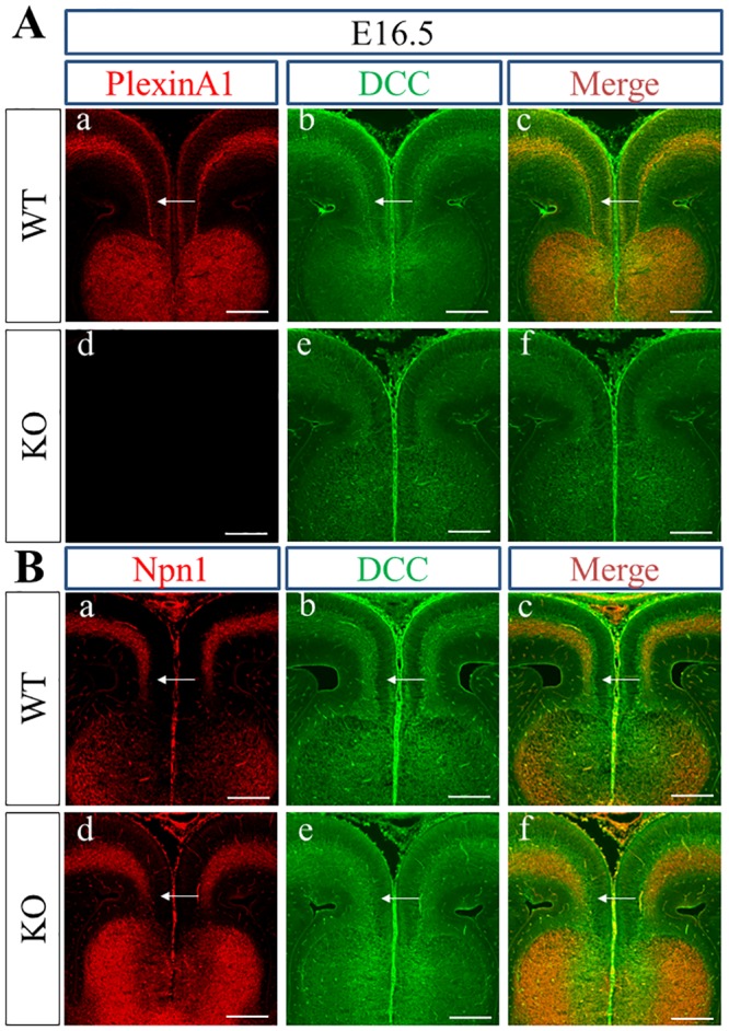
Fig 2. Localization of PlexinA1, DCC, and Npn1 in WT and PlexinA1 KO brains at E16.5.

(A) PlexinA1 (red) was expressed in the DCC+ area (green) deep within the cingulate cortex in WT brains at E16.5 (arrows in a, b, c), but only DCC (green) was detected deep within the cingulate cortex in PlexinA1 KO brains (d, e, f). (B) Expression of Npn1 (red) and DCC (green) overlapped with the DCC+ area (green) in WT (arrows in a, b, c) and PlexinA1 KO (arrows in d, e, f) mouse brains at E16.5. Scale bars: 200 μm.
