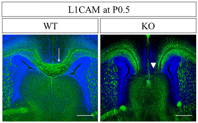
Fig 9. Agenesis of CC in PlexinA1 KO mice at P0.5.
Coronal brain sections of WT and PlexinA1 KO mice at P0.5 were stained with anti-L1CAM antibody in the middle levels of CC. L1CAM+ callosal axons crossed the cortical midline in 16 out of 16 WT mice (arrow in WT). In contrast, L1CAM+ callosal axons did not cross the midline in 10 out of 13 PlexinA1 KO mice (arrowheads in KO). Scale bars: 200 μm.

