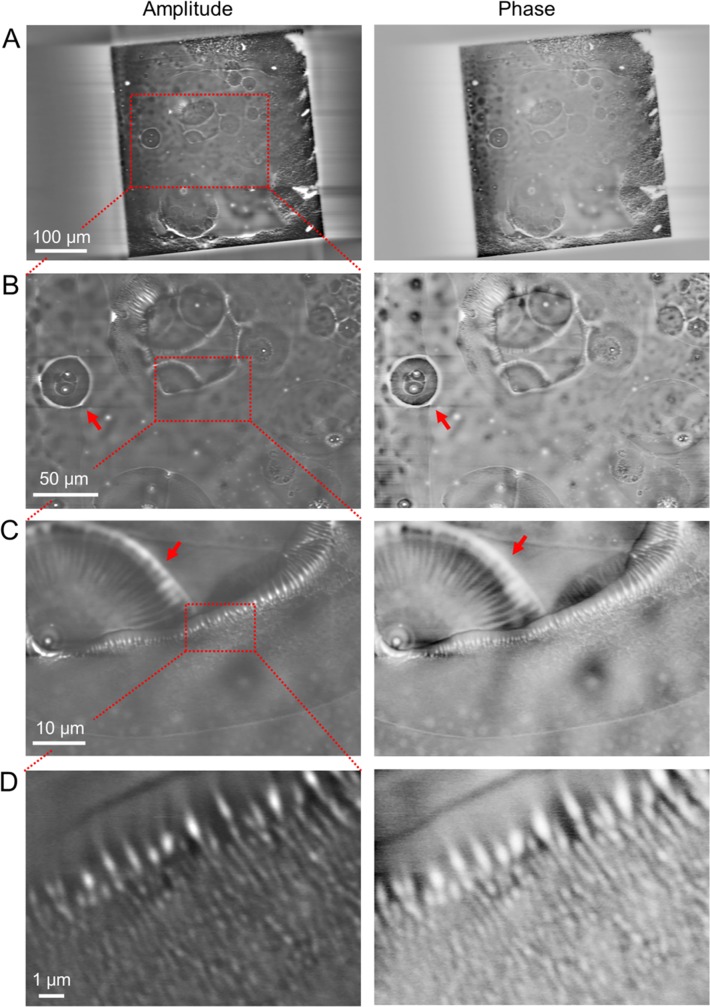
Fig 4. Amplitude and phase images of PLA beads in the bubble area of a wet environment observed by the IP-SEM system.
(A) Amplitude and phase images of the 500 nm-diameter PLA beads in water with bubbles (EB accelerating voltage 5 kV, EB current 400 pA, input frequency 50 kHz, magnification 200×). Large spherical bubble areas are clarified in both images. (B) Expanded amplitude and phase images of the red boxed area in (A) at 500× magnification. Both images clarify the white contrast of aggregation beads and the spherical bubble areas. (C) Amplitude and phase images of the air–water boundary of the bubble in the red boxed area of (B) at high-magnification (2,000×). (D) High-magnification (10,000×) amplitude and phase images of the air–water boundary of the bubble. Both images in (C) and (D) clarify the white-contrasted beads along the bubble boundary. These amplitude images were reversed contrast. Scale bars: 100 μm in (A), 50 μm in (B), 10 μm in (C) and 1 μm in (D).

