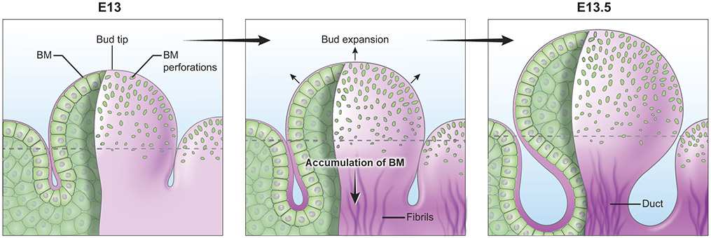
Figure 5. BM micro-perforations in embryonic mouse submandibular salivary gland.
Micro-perforations are prominent at the tip of epithelial buds. These perforations gradually disappear towards the equator (dotted line). As the growing buds expand rapidly, the number of perforations peaks at embryonic day (E) 13.5. Basement membrane translocation away from the bud tip is accompanied by accumulation of fibril-like basement membrane proteins at the forming secondary duct.

