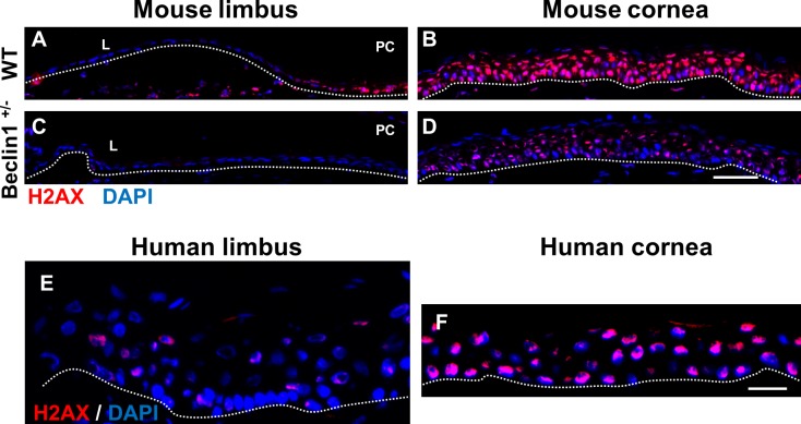
Figure 8.
scRNA-seq identifies a reduction in H2ax expression in beclin 1+/− mouse corneal epithelial cells. (A–D) Immunofluorescence staining with H2AX antibody in a frozen section of mouse limbal and corneal epithelium. In wild-type mice, H2AX is preferentially expressed in the corneal epithelium (B) compared with limbal epithelium (A). H2AX expression is significantly decreased in the corneal epithelium from beclin 1+/− mice (D). n = 4. Scale bar: 50 μm. (E, F) Immunofluorescence staining with H2AX antibody in a frozen section of human limbal (F) and corneal (E) epithelium. H2AX was primarily restricted to the corneal epithelium. Scale bar: 20 μm. Dotted lines mark the basement membrane. n = 3.

