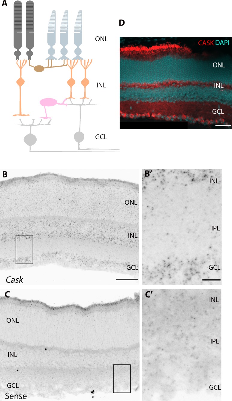
Figure 3.
CASK is expressed in RGCs. (A) Schematic of mouse retina. ONL, outer nuclear layer; INL, inner nuclear layer; GCL, ganglion cell layer. (B) In situ hybridization shows Cask mRNA is expressed in the INL and GCL of the P14 retina. Scale bar: 50 μm. (B') shows higher magnification of expression in GCL. Scale bar: 10 μm. (C) No appreciable signal was detected in the GCL with the sense riboprobe. (C') shows higher magnification of GCL in C. (D) CASK immunoreactivity in the GCL of P21 retina. Scale bar: 50 μm.

