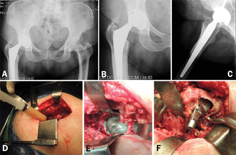
Fig. 1.

Asymptomatic patient submitted to total hip arthroplasty 4 years ago. Radiographically we observe the excessive wear of the polyethylene, not compatible with the period of service of the implants. ( Fig 1. A–C ). We performed revision surgery despite the absence of clinical manifestations or tests indicative of infection. Preoperative tests: ESR = 19mm, CRP = 29.2 mg/L, dimer D = 530 ng/mL Intraoperative aspiration of the hip revealed an abundant amount of purulent-looking liquid. ( Fig. 1-D ) We could not observe any signs of infected periprosthetic tissues, acetabular loosening or third body abrasion. ( Fig. 1-E ) Large area of osteolysis was observed in the posteromedial region of the proximal femur, which extended to the trochanteric region. After curetting the whitish and friable tissue, an extensive area of bone loss could be seen in the proximal femur. ( Fig. 1-F ) Intraoperative tests: Leukocyte esterase: +; Synovial leukocytes: 52,800; % Neutrophils: 50%. All cultures of periprosthetic tissue and 1 culture of synovial fluid (in blood culture medium) were negative after 8 days of incubation. Up to 18 months postoperatively, the patient was asymptomatic and without any changes in the serum markers for infection.
