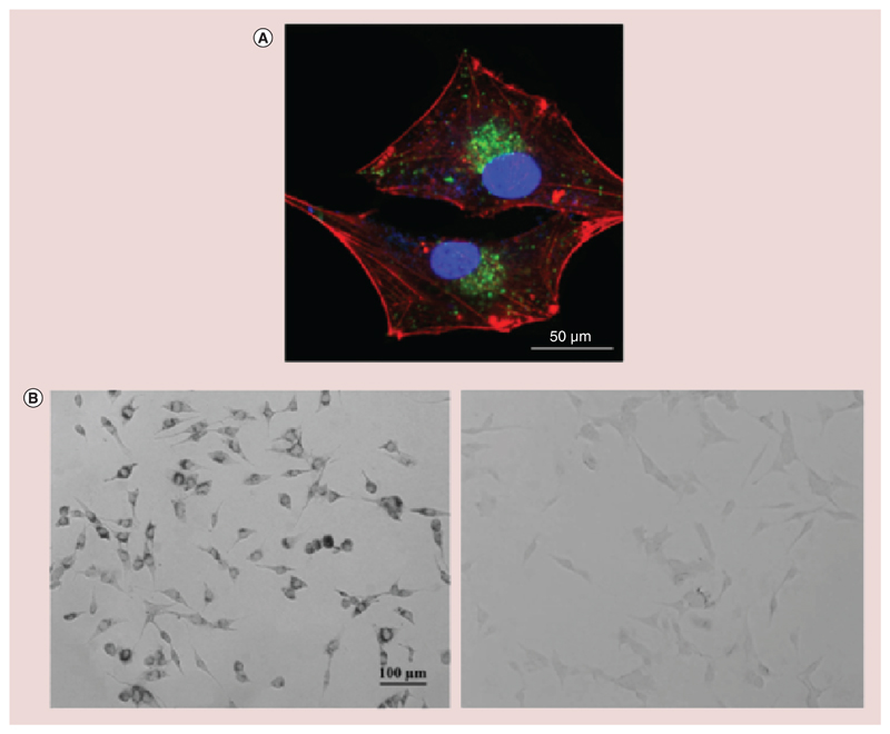
Figure 4. Cellular uptake investigation.
(A) Representative confocal fluorescence image of U-87 MG cells showing FITC-Nut-Mag-SLNs uptake after 24 h of incubation (nanoparticles in green, f-actin in red, nuclei in blue). (B) Prussian blue staining of U-87 MG cells after 24 h of incubation with Nut-Mag-SLNs (on the left). The image shows blue precipitates in the perinuclear area due to the presence of SPIONs upon Nut-Mag-SLN internalization. Nontreated cells are reported as control (on the right).
Nut-Mag-SNLs: Nutlin-loaded magnetic solid lipid nanoparticles; SPIONs: Superparamagnetic iron oxide nanoparticles.

