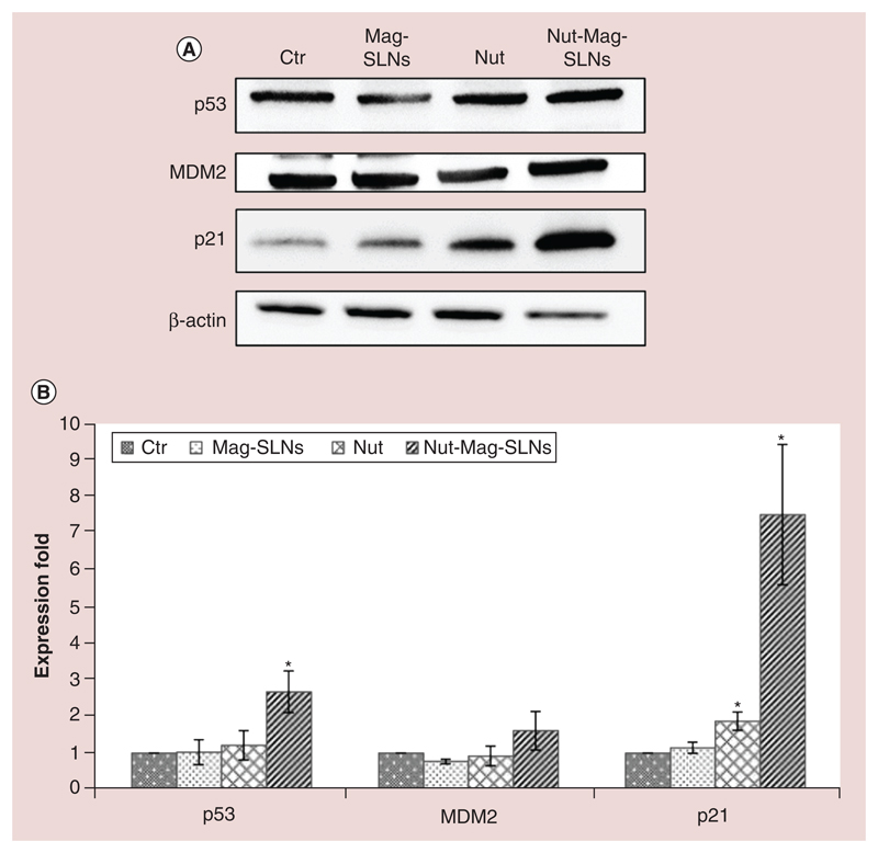
Figure 6. Western blotting analysis of markers involved in apoptosis.
(A) Expression of p53 and its downstream proteins (MDM2 and p21) was analyzed on U-87 MG cells after 72 h of treatment with 100 μg ml-1 of Mag-SLNs, 1.33 μM of nutlin-3a, or 100 μg ml-1 of Nut-Mag-SLNs (corresponding to 1.33 μM of drug), and compared with control cultures. (B) Quantitative evaluation of western blotting results.
*p < 0.01.
MDM2: Murine double minute; Mag-SLNs: Magnetic solid lipid nanoparticles; Nut-Mag-SNLs: Nutlin-loaded magnetic solid lipid nanoparticles.

