Figure 2.
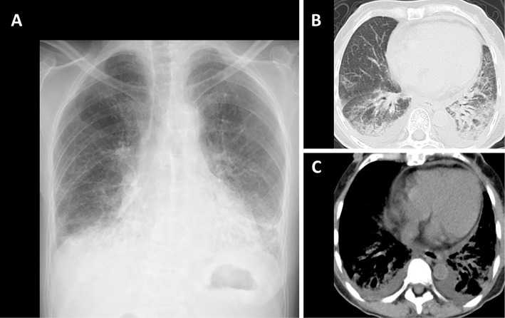
Chest radiography findings at the initial presentation. Ground-glass shadows are observed in the bilateral lower lung fields (A). Chest CT shows ground-glass opacities and consolidations in the bilateral lower lobes (B) and small volumes of bilateral pleural effusion and pericardial effusion (C).

