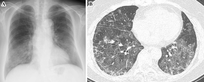
Figure 1.
(A) Chest radiography at hospitalization (late December 2015) shows diffuse infiltrate shadows in the bilateral lower lung fields. (B) Chest high resolution computed tomography at hospitalization shows bilateral ground glass opacities and intralobular and interlobular septal thickening, which were most prominent in the lower lobes.

