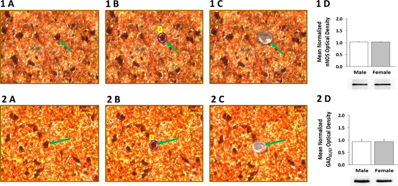
Figure 1. Laser-Catapult Microdissection of Immunolabeled Ventromedial Hypothalamic Nucleus (VMN) Nitrergic- or γ-Aminobutyric Acid (GABA) Neurons: Western Blot Confirmation of Accuracy of Immunocytochemical Identification of Neurotransmitter Phenotype.
VMN neurons were identified in situ for neuronal nitric oxide (nNOS)- [top row; Panel 1 A] or glutamate decarboxylase65/67 (GAD65/67)-[bottom row; Panel 2 A] immunoreactivity (-ir); representative nNOS- or GAD65/67-ir-positive neurons is indicated by green arrows. Areas shown in Panel 1 A and 2 A were re-photographed after positioning of a continuous laser track (depicted in blue) around a single nNOS-ir [Panel 1 B; green dashed arrow] or GAD65/67-ir neuron [Panel 2 B; green dashed arrow] and subsequent ejection of that cell by laser pulse [Panels 1 C and 2 C]. Note that this microdissection technique causes negligible destruction of surrounding tissue and minimal inclusion of adjacent tissue. Panels 1 D and 2 D show that nNOS or GAD65/67 protein is expressed in pure VMN nerve cell samples identified immunocytochemically for nNOS or GAD immunoreactivity, respectively.

