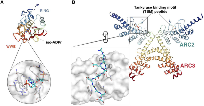
Figure 3. Structures of the RNF146-iso-ADPr complex and ankyrin repeat clusters (ARCs) of PARP5a binding to Tankyrase binding motif (TBM) of RNF146.

(A) Cartoon model of the RNF146- iso-ADPr complex highlighting the RING finger and WWE domain (PDB ID: 4QPL), with a close-up of the RNF146-iso-ADPr binding interface. (B) Cartoon model of the complex between RNF146 TBM and PARP5a (PDB ID: 6CF6), with a close-up of the TBM binding interface at an ARC of PARP5a. Structures were generated via Protein Data Bank and PyMOL.
