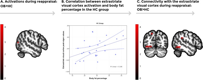
Fig. 2.
a Brain regions showing increased activation in the obese group during the emotion regulation task (Regulate vs. Maintain). Increased activation was found in the right extrastriatal visual cortex in the obese group in comparison to the HC group (p < 0.05, AlphaSim cluster-extent corrected). Color bar represents t-values. b A scatterplot depicting the positive correlation between extracted activation eigenvalues from the extrastriatal visual cortex peak during the emotion regulation task (Regulate vs. Maintain) and total body fat percentage in the control group [n = 25, (r(23) = 0.431, p = 0.031)]. Error bars show the 95% confidence interval (CI). c Increased psychophysiological interactions (PPI) connectivity using the extrastriatal visual cortex as a seed was found in the left inferior temporal lobe, the left precuneus, and the left supramarginal gyrus in the obese group in comparison to the healthy control group. Color bar represents t-values

