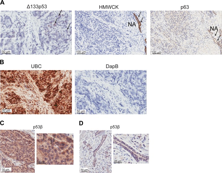
Fig. 3. Δ133TP53β is expressed in cancer cells.
a In situ hybridization using RNAscope detected Δ133TP53 (black arrows) in FFPE prostate cancer tissues (left panel). Immunohistochemical staining for high molecular weight cytokeratin (HMWCK) and p63 (middle and right panels, respectively) to identify loss of HMWCK and p63 in prostate cancer. NA normal associated tissue (black arrows). b Left panel, probes to ubiquitin C (UBC) as a positive control for RNA quality and right panel, probes to the bacterial gene DapB as a negative control. Nuclei were counterstained with hematoxylin. c Immunohistochemistry using the KJC8 antibody to detect p53β (black arrows) in FFPE prostate cancer tissues. d Absence of p53β staining in normal associated prostate epithelium. Nuclei were counterstained with hematoxylin

