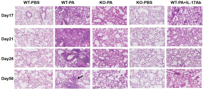
Figure 4.
Representative images of HandE staining of the WT-PBS, WT-PA, KO-PA, KO-PBS, and WT-PA + IL-17Ab groups. Lung tissue specimens from the five groups (n = 6 each group) were collected on the 17th, 21st, 28th, and 56th day and stained. The KO-PA and WT-PA + IL-17Ab groups show less inflammatory cell infiltration on the 17th, 21st, 28th day than the WT-PA group and no loose granuloma on the 56th day.

