
FIGURE 5.
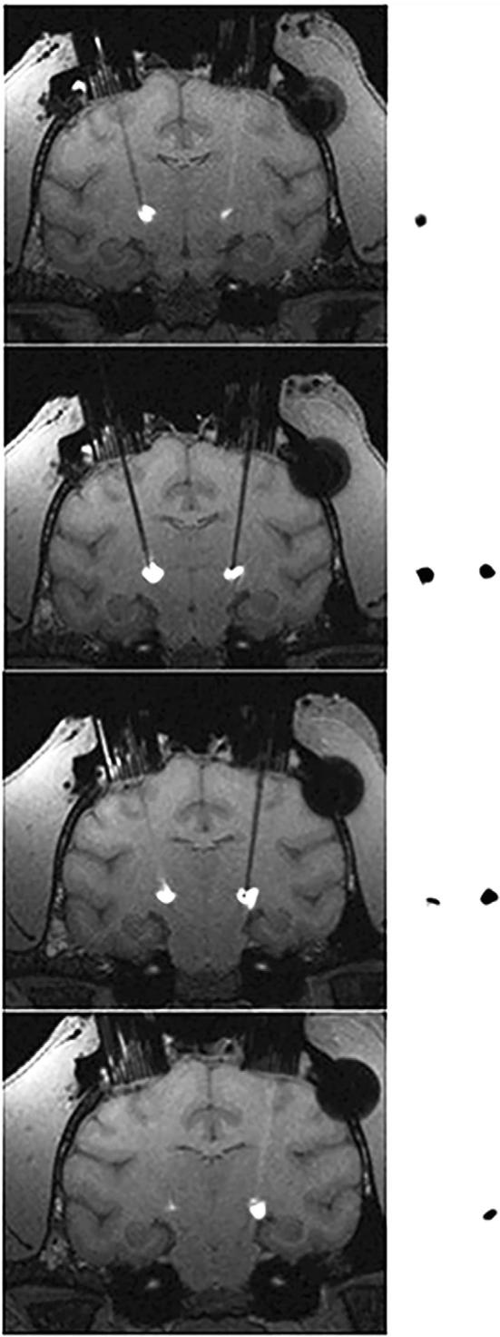
Comparison between T1-weighted MRI and autoradiograms. Representative serial coronal slices of T1-weighted MR images post-contrast were compared to corresponding autoradiographic sections (20 μm) from an NHP who underwent co-infusion of 3H-muscimol and Gd-DTPA. Anatomically registered MRI-autoradiogram pairs are separated by 1 mm.