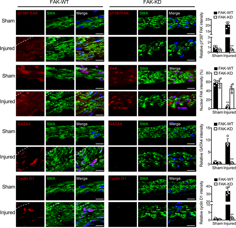
Figure 7. SMC-specific FAK-KD mice develop reduced hyperplasia and exhibit less GATA4 and cyclin D1 expression after wire injury.
Immunofluorescence staining of FAK-WT and FAK-KD femoral arteries 4 weeks postinjury for pY397 FAK, FAK, GATA4, cyclin D1, and a-SMA. Red, green (α-SMA), and blue (DAPI) were merged. Relative fluorescence intensity of nuclear FAK and indicated targets in a-SMA positive cells were quantified (±SD; n=5). Scale bars, 10 μm. Dashed lines, boundary between media and intima. **P<0.005

