
Fig. 4.
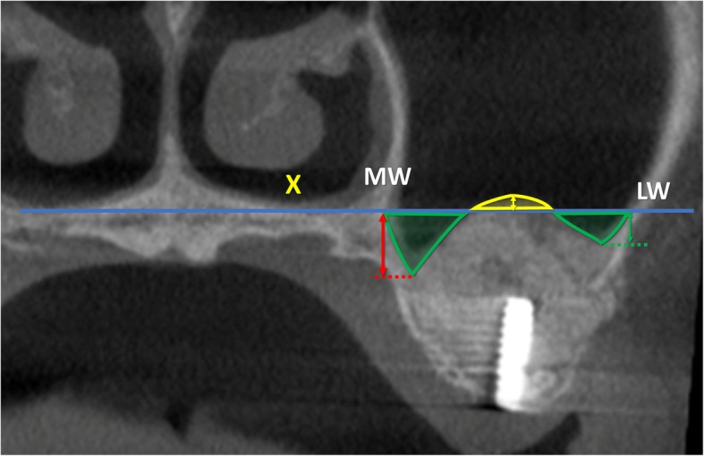
T2, coronal view of a no-membrane CBCT; X, line drawn following the floor of the nose; MW, medial wall of the sinus; LW, lateral wall of the sinus; Exceeding area (bordered in yellow), area above the line X filled with biomaterial/ bone tissue. Residual areas (bordered in green), the area below the line X not filled with biomaterial/ bone tissue. Floor augmentation heights at the lateral (green arrow), middle (yellow arrow), and medial (red arrow) aspects