
Fig. 19.
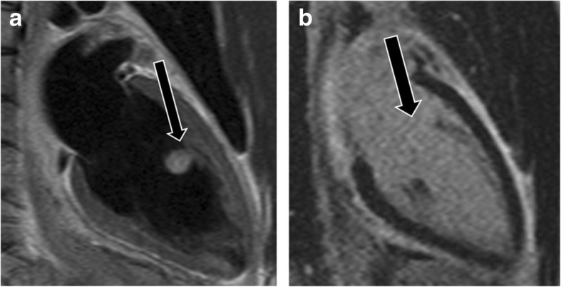
Myxoma. a Two-chamber T2-weighted dark blood image shows a hyperintense mass involving the anterolateral papillary muscle (arrow). b The mass enhances after contrast-enhancement (arrow) making it isointense to the blood pool. This was proven to be a myxoma