Figure 3.
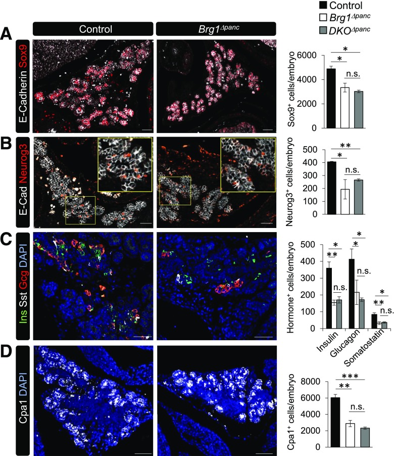
All pancreatic cell lineages are reduced in e15.5 Brg1Δpanc and DKOΔpanc epithelium. Control, Brg1Δpanc, and DKOΔpanc mutant embryos were stained with antibodies specific for E-cadherin and Sox9 (A), E-cadherin and Neurog3 (B), insulin (Ins), somatostatin (Sst), and glucagon (Gcg) (C), or Cpa1 (D). The yellow square in B marks the magnified area in the panel. DAPI nuclear staining is also provided in C and D. Cell type counting was performed on sections 90-µm apart that spanned the entire pancreatic region (n = 3). *P < 0.05. **P < 0.01. ***P < 0.001. n.s., not significant. Scale bar = 50 µm.

