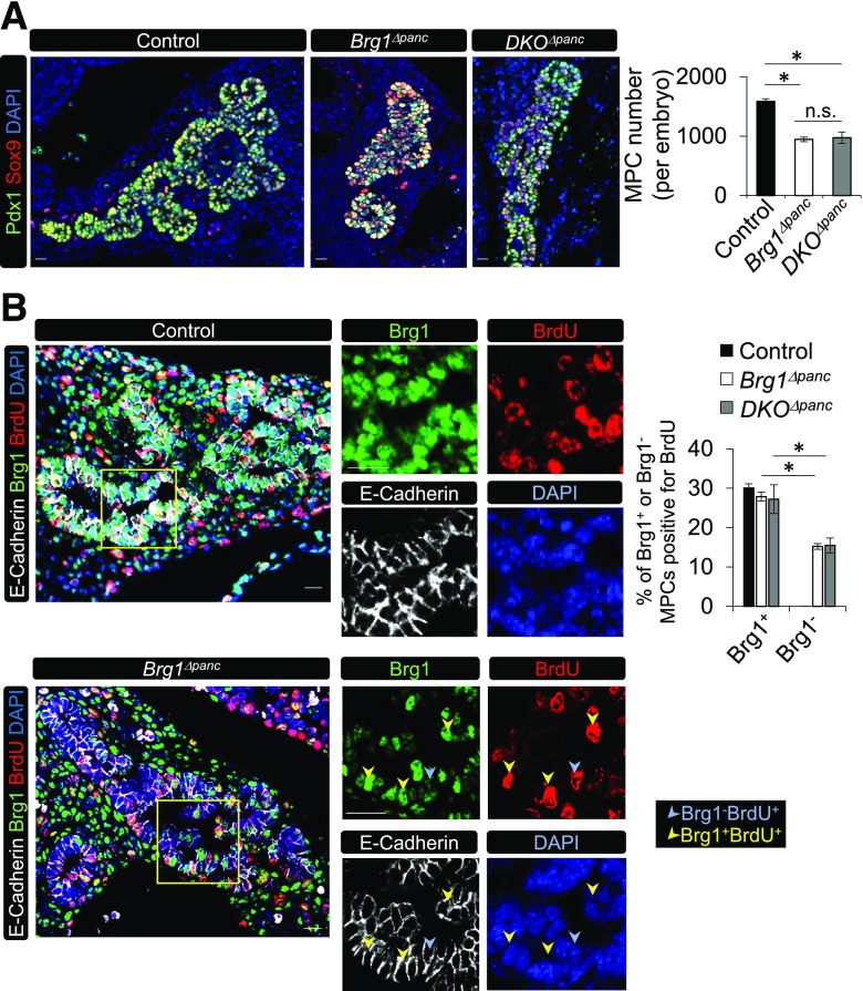
Figure 4.
MPC proliferation is dependent on the Brg1 ATPase subunit of Swi/Snf. A: A representative immunostaining image of Sox9+ and Pdx1+ cells in control, Brg1Δpanc, and DKOΔpanc pancreata at e12.5. Sox9+Pdx1+ MPC numbers were determined in sections obtained every 60 µm of the entire pancreatic region. B: E-cadherin, Brg1, and BrdU staining of pregnant dams injected with BrdU 30 min before the e12.5 embryos were harvested. Also provided is the percentage of Brg1+ or Brg1− cells that incorporated BrdU (n = 3). The yellow square illustrates the magnified area displayed in the panels on the right. *P < 0.05. Scale bar = 20 μm. DAPI nuclear staining is shown in A and B.

