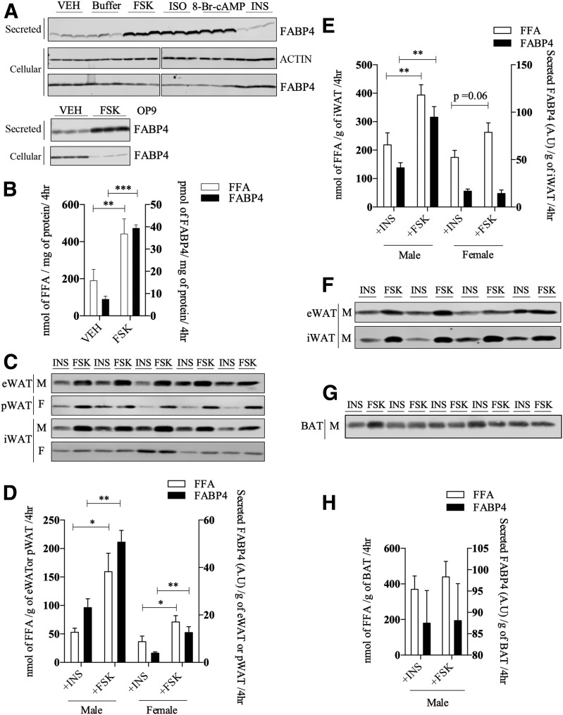
Figure 1.
FABP4 secretion in response to lipolytic stimuli. A (top): Western analysis of FABP4 secretion from differentiated 3T3-L1 adipocytes in response to a 4-h treatment with VEH, buffer, 20 μmol/L FSK, 10 μmol/L isoproterenol (ISO), 1 mmol/L 8-Br-cAMP, or 500 nmol/L insulin (INS). The intracellular levels of β-actin and FABP4 were determined immunochemically. Because these samples were analyzed on different gels, the indicated break in the image denotes the two separate gels used. A (bottom): Secreted and intracellular FABP4 in differentiated OP9 adipocytes in response to VEH or 20 μmol/L FSK. B: Levels of FABP4 (quantified from A) and fatty acids (FFA) secreted (as measured by the colorometric assay described in research design and methods) by 3T3-L1 adipocytes treated with VEH or 20 μmol/L FSK. C: Tissue explants from eWAT, pWAT, and iWAT were isolated and treated with 20 μmol/L FSK or 500 nmol/L INS for 4 h, and the secretion of FABP4 was evaluated immunochemically. D: Quantitation of FABP4 (from C) and FFA secretion by eWAT and pWAT explants. E: Quantitation of FABP4 and FFA secretion by iWAT explants from male or female C57BL/6J mice as shown in C. F: FABP4 secretion from primary adipocytes derived from eWAT and iWAT of high-fat–fed C57BL/6J mice in response to 2-h treatment with either 20 μmol/L FSK or 500 nmol/L INS. G: Explant tissue from interscapular BAT of male C57BL/6J mice was isolated as described and treated with 20 μmol/L FSK or 500 nmol/L INS for 4 h. The secretion of FABP4 was evaluated immunochemically. H: Quantitation of FABP4 and FFA secretion by BAT explants from male C57BL/6J mice as shown in G. *P < 0.05; **P < 0.01; ***P < 0.001. A.U., arbitrary units; F, female; hr, hours; M, male.

